How the Eye Detects Light
For us to see light, the eye must first convert light into an electrical signal that the brain can understand. This begins in the retina, which contains millions of light-sensitive cells called rods and cones. Inside these cells is a tiny light-sensitive molecule called retinal, held within a much larger protein called opsin.
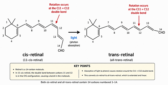
When a particle of light, called a photon, is absorbed by retinal, its energy causes the retinal molecule to change shape—from a bent shape to a straighter one.
Because retinal fits tightly inside the opsin protein, this change pushes against the surrounding protein and causes the opsin itself to change shape.
That change acts like flipping a microscopic switch. It starts a series of chemical reactions inside the cell that ultimately changes the flow of electrically charged particles across the cell membrane. This sends the electrical signal that light has been detected.

More Detail
Retinal is able to change shape when it absorbs light because of the unusual properties of the double bonds that connect the carbon atoms in its backbone.
To understand this, it helps first to picture what an electron orbital actually is. An electron does not orbit an atomic nucleus like the Moon orbits the Earth. Instead, it occupies a region of space around the nucleus where it is likely to be found. These regions are called orbitals, and they can be thought of as electron clouds with particular shapes.
Retinal contains a chain of carbon atoms connected by alternating single and double bonds. At each carbon involved in a double bond, three of its bonds lie in a flat plane and point in directions about 120 degrees apart. You can picture them as pointing toward the 4, 8, and 12 o'clock positions on a clock face, with the carbon nucleus at the center.
This arrangement gives the carbon chain its zigzag shape. When successive parts of the chain bend in opposite directions, the bends largely cancel and the chain remains relatively straight. But when neighboring parts bend in the same direction, a pronounced bend or kink is produced. This is what gives the cis form of retinal its characteristic bent shape.
A carbon-carbon double bond also has another important feature. Above and below the flat plane of the carbon atoms are electron clouds that overlap with one another. Together they form what is called a pi (π) bond. You can picture these overlapping clouds as forming an hourglass-like region above and below the carbon-carbon bond.
The π bond acts like a lock, preventing the carbon chain from rotating around the double bond. This holds retinal in its bent cis form, even though the straighter trans form is more stable. The cis form is somewhat like a spring being held under tension.
The chain cannot rotate around the double bond until a photon arrives with just the right amount of energy. The photon transfers its energy to an electron in the π bond, lifting it into a higher-energy state and briefly breaking the π part of the double bond. For an extraordinarily brief moment, the lock is released, allowing the carbon chain to rotate and snap into the more stable trans shape.
The change happens extraordinarily quickly, in a matter of femtoseconds.